
Laser Hair Removal Hypopigmentation: A Complete Guide
You've booked laser hair removal before, or you're thinking about it now, because you want smoother skin without the daily hassle. Then you hear the word hypopigmentation, and the conversation suddenly feels less simple. A few lighter patches after treatment can be confusing, especially if they show up weeks later and you can't tell whether your skin is healing normally or reacting to the laser.
Laser hair removal hypopigmentation means a loss of pigment that makes part of the treated skin look lighter than the skin around it. It's different from hyperpigmentation, which is darkening. If you've noticed flat pale patches on the legs, underarms, bikini line, face, or neck after a session, you're not overreacting, and you're not alone in wanting a straight answer about what it is, why it happens, and whether it goes away.
The good news is that pigment loss after laser hair removal isn't mysterious once you understand how lasers and skin pigment interact. A better question is not just, “Can this happen?” but “How do I lower the odds, spot it early, and choose a clinic that plans for diverse skin tones from the start?” For readers in Long Island and Nassau County, that question matters even more when Fitzpatrick IV to VI skin is in the picture and the device choice can change the risk conversation.
When a Smooth Result Comes with Pale Patches
A client leaves the office feeling relieved because the hair is shedding and the skin looks settled, so the appointment seems to have done exactly what it promised. Then, a few weeks later, she notices a pale patch near the bikini line or underarm in the mirror. It is not raised, it does not itch, and it can look like the skin is still healing, so she waits and hopes the color will blend back in on its own.
That waiting period is where a lot of confusion begins. Hypopigmentation is a reduction in melanin, the pigment that gives skin its color, so the treated area looks lighter than the skin around it. It is different from a scab, a rash, or leftover redness, and it is also different from the brown discoloration people usually expect after skin injury.
What it tends to look like
Post-laser pale patches are often flat, non-itchy, and found right in or near the treated zone. The border can be sharp or soft, depending on skin tone, how much energy was used, and how the skin responded. People often mistake early pigment loss for incomplete healing because the surface can look intact even while the pigment-making cells have been stressed.
The clinical picture is not rare enough to ignore, especially in people with darker skin tones. A careful clinic should be able to explain how the laser choice, skin type, and cooling strategy all fit together, since that is the difference between a treatment plan that removes hair and one that also respects pigment balance. If you want to see how providers describe that broader approach, advanced laser technology explained by NY Laser and hair removal from Chernoff Cosmetic Surgery are useful places to compare how clinics talk about safety, skin selection, and treatment goals.
Practical rule: pale patches that appear after laser hair removal deserve observation, not panic, but they also deserve a documented follow-up plan.
The next sections cover why these patches form, who is most vulnerable, and what helps color return.
How Lasers Interact with Skin Pigment
Hair-removal lasers are designed to find melanin in the follicle and heat it enough to damage the hair unit. That's the goal. The problem starts when some of that heat reaches the surrounding epidermis and injures the melanocytes, the cells that make pigment in the skin.
A magnifying glass focuses sunlight on one spot. If the beam is well matched to the target, the heat stays where it should. If the settings are too aggressive, the wavelength is poorly chosen, or cooling isn't enough, the energy spreads into nearby tissue and the pigment cells take the hit.
Why wavelength matters
The two wavelength families frequently mentioned are alexandrite 755 nm and Nd:YAG 1064 nm. In plain language, shorter wavelengths are absorbed more readily by epidermal melanin, while the longer Nd:YAG wavelength tends to bypass more of the surface pigment, which is why it's generally considered safer for darker skin. That doesn't make any device automatically safe, but it explains why matching wavelength to skin tone matters so much.
A Long Island patient comparing device types may find a general overview like what to expect with laser hair removal helpful for understanding the basic process before the consult. For more advanced device context, an internal resource on advanced laser technology can help frame why platform choice affects safety and comfort.
Three settings shape the risk conversation most directly:
Fluence, the amount of energy delivered, because higher energy raises the chance of thermal injury.
Pulse duration, which is how long the energy is delivered, because heat spread changes with timing.
Epidermal cooling, which protects the surface skin by pulling heat away from the skin layer that shouldn't be overtreated.
Plain-language definition: reversible melanocyte suppression means the pigment cells are stressed but recover over time, while permanent suppression means the cells are destroyed or never fully return to normal activity.
That distinction is the difference between a patch that fades and a patch that lingers.
Who Is Most at Risk and Why
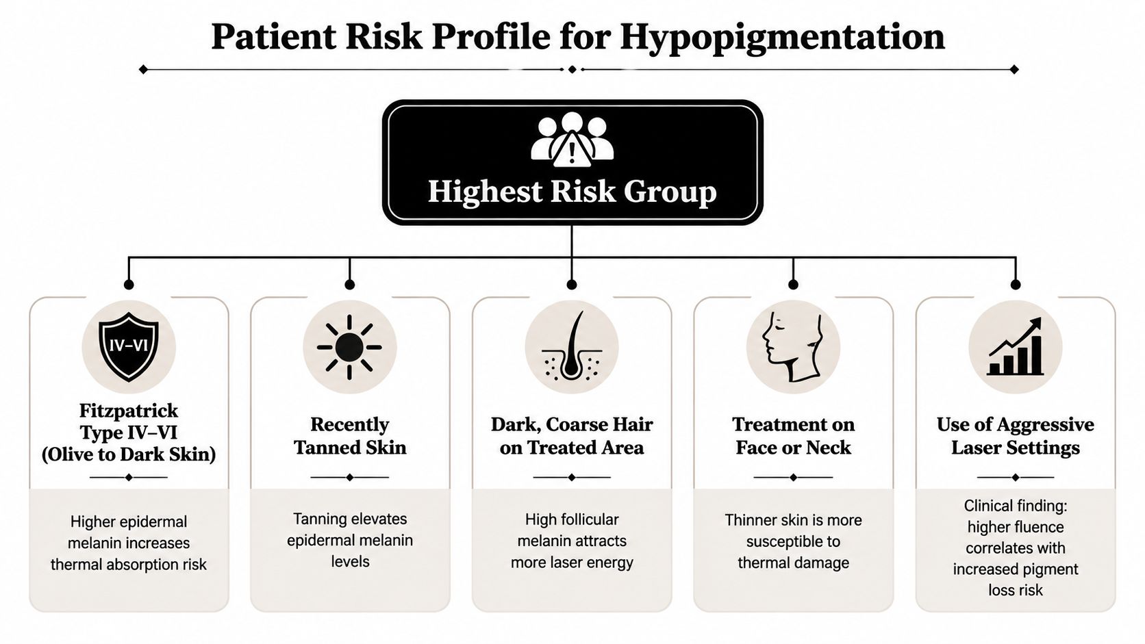
A first-time patient often asks a simple question before the chair is even reclined, who is most likely to end up with pale patches after treatment? The answer depends less on the word “laser” itself and more on how much pigment sits in the skin around the follicle, how reactive that skin is, and how carefully the treatment is matched to the patient's tone. Skin that already carries more melanin, especially Fitzpatrick IV to VI, needs a more cautious plan because the margin for error is smaller.
The combinations that raise concern
Risk rises when several variables point in the same direction. A dark skin type treated with aggressive settings is one example. Recently tanned skin is another, because fresh pigment in the epidermis gives heat more target to absorb. If a provider is not adjusting for that extra pigment, the skin can be treated as if it were lighter than it really is.
Areas with thinner or more reactive skin can also show pigment change more easily. The underarm, shin, face, and neck often leave less room for heat to dissipate safely than thicker body areas. A patient who has already had color changes after inflammation, acne, waxing, or other irritation may be more likely to see a visible contrast after laser treatment, even when the session seems routine.
This is why a provider should ask about the whole skin story, not just hair growth. A clinic that regularly treats darker phototypes should know how to adapt device choice, cooling, and fluence for that population, and patients can use a local resource like laser hair removal dark skin near me to compare whether a practice appears set up for Fitzpatrick IV to VI care.
The practical concern is not only whether the laser removes hair, but whether the skin around it stays evenly colored while it heals. Readers who want a broader picture of long-term concerns can review pigment changes from laser hair removal as a reminder that pigment change belongs in the consult, not in the afterthought pile.
A quick self-check
Cautious tier: dark skin, recent tan, or prior pigment changes.
Moderate tier: medium skin with a history of easy irritation or inconsistent sun protection.
Lower-risk tier: untanned skin, a device chosen for the skin tone, conservative settings, and no prior pigment issues.
The point is not to frighten anyone away from treatment. The point is to show that risk is modifiable, and that a careful provider should adjust the plan before the first pulse is fired.
Pre-Treatment Habits That Prevent Pigment Loss
Prevention starts before the machine is ever turned on. If you arrive with tanned skin, irritated skin, or a hidden medication issue, the skin is already at a disadvantage. Good prep lowers the chance that heat will spread where it shouldn't.
The four controls that matter most
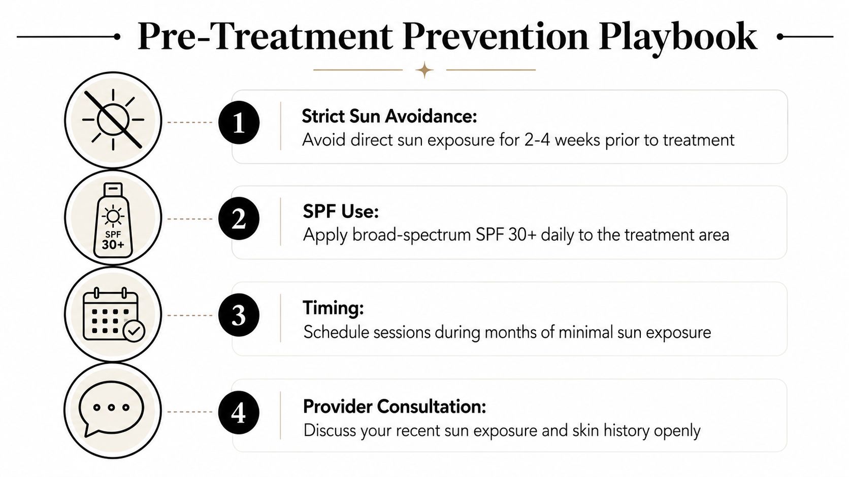
A careful provider will usually focus on four things first. Sun avoidance and SPF use matter because recent tanning increases epidermal melanin and makes selective heating harder. Patch testing on a discreet area helps show how the skin responds before treating a larger zone.
The next two are device-centered. Conservative fluence means starting with lower energy and increasing only if the skin tolerates it. Strong epidermal cooling matters because it protects the surface layers while the follicle is being targeted, which is the whole point of reducing non-selective heat.
Patient behavior matters too. If you're using retinoids, exfoliants, or recent peel products, tell the provider before the session. Those products can make skin more reactive, even when they're not the direct cause of pigment loss. A full history includes medications, recent cosmetic procedures, and any prior blistering or color change.
Questions worth asking before treatment
Patch test plan: Will they test a small area first if your skin tone or tanning history raises concern?
Setting strategy: Do they start conservatively and adjust based on the skin's response?
Cooling method: What cooling is built into the device, and how do they confirm it's working?
Sun exposure check: Are they asking about recent tanning, outdoor sports, or vacation sun before booking?
The most predictive prevention behavior is still choosing a wavelength that matches the skin type. That's why device selection matters so much for darker skin, and why technology discussions belong in the consult rather than being left until something goes wrong.
If you want a visual refresher on how providers think through patch assessment, the embedded timeline above pairs well with the clinical question of whether a spot is healing or shifting in a more concerning direction.
Diagnosing the Patches and Reading the Timeline
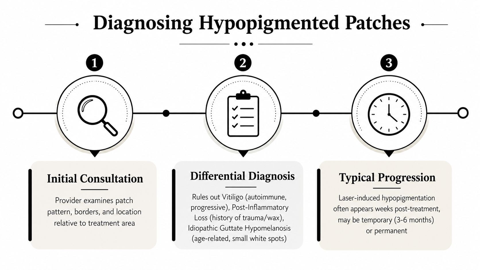
A pale patch after treatment does not automatically mean laser injury, and a careful clinician should show why before naming the cause. The first clue is the history. If the lighter area appeared after a laser session and stays exactly inside the treated zone, that points in one direction. If it was already present, spreads beyond the hair-removal field, or shows up with other white patches elsewhere, the diagnosis needs a wider look.
Wood's lamp examination is often used because it can make pigment differences easier to see. In stubborn cases, a biopsy may be considered, especially if the borders are expanding or another diagnosis is still possible. The goal is to separate laser-induced hypopigmentation from conditions like vitiligo, post-inflammatory pigment loss, or idiopathic guttate hypomelanosis, which can look similar at first glance but behave very differently.
What the timeline usually tells you
The timeline can be reassuring, but only if you read it carefully. Mild cases may start repigmenting within weeks. Some cases can last for about two months, and a case report documented a single-session injury that fully recovered by 12 months with conservative care and sun protection. A long-pulsed alexandrite series also described reversible melanocyte suppression with gradual return toward baseline color over time (PubMed 23384006).
That is why a pale patch is not automatically permanent just because it is still visible at the next appointment. The follow-up window should be measured in months, not days. In that same clinical summary, long-pulsed alexandrite photo-epilation was associated with overall complications in 2.2% of cases, while transient hypopigmentation accounted for 0.5% and no long-term pigmentary change appeared in that series.
A simple photo log helps more than many people expect. Take pictures in the same light every one to two weeks, and keep the angle, distance, and background consistent. Those comparisons help the provider see whether the borders are fading, staying flat, or expanding.
Sun protection is not just a comfort measure during recovery. It is part of the diagnostic process, because UV exposure can delay repigmentation and make the timeline harder to read.
If the patch is stable but not improving, that does not mean panic. It means the clinician needs a better look at the pattern, the skin history, and whether the original laser settings still make sense. For a closer look at what recovery monitoring can involve, see the guide on after-laser treatment for pigmentation.
Evidence-Based Options for Bringing Color Back
Treatment should match the pattern, the duration, and the severity. The mildest cases often need nothing more than time, strict photoprotection, and a skin-care routine that doesn't irritate the area further. That sounds simple, but it's often the right answer when pigment loss is temporary and the skin is otherwise quiet.
A stepped treatment menu
At the conservative end, clinicians may recommend sun protection, gentle moisturization, and waiting for repigmentation. Some practices also use topical antioxidants such as vitamin C as supportive care, especially when the skin is still settling. These aren't dramatic fixes, but they suit stable patches that are already recovering.
More active topical options show up when pigment loss lingers. Clinical discussions of post-inflammatory hypopigmentation include tacrolimus, psoralen plus UVA, and melanocyte-stimulating approaches. For small stable patches, targeted office treatments such as narrowband UVB or 308 nm excimer laser are often considered because they aim light directly at the pale area rather than treating the whole field.
Combination approaches may be used when the skin needs a stronger push. That can include microneedling-based strategies, sometimes paired with platelet-rich plasma or other adjuncts, with the goal of nudging pigment cells and their supporting environment back toward normal activity. For persistent or spreading patches, referral to dermatology is appropriate, especially if the borders suggest vitiligo or the patient is distressed by the appearance.
The internal article on after laser treatment for pigmentation is a useful local reference for readers who want to understand how post-treatment skin changes are approached in a clinical setting.
Comparing Hypopigmentation Treatment Options
Option | Best For | Evidence Note | Typical Setting |
|---|---|---|---|
Photoprotection and time | Fresh or slowly improving patches | First-line conservative care | Home care |
Topical support, including tacrolimus or vitamin-based support | Mild persistent color loss | Used in stepwise clinical practice | Home care with follow-up |
Narrowband UVB | Localized stable patches | Common in pigment recovery protocols | Dermatology office |
308 nm excimer laser | Small focal white patches | Used for targeted repigmentation | Dermatology office |
Referral and broader workup | Expanding or unclear lesions | Needed when diagnosis is uncertain | Dermatology evaluation |
One case series noted that pigment can decline again around 6 months after excimer therapy, which is why follow-up matters even after visible improvement. The point isn't that every patient needs every treatment. It's that persistent white patches deserve a plan, not reassurance alone.
What to Ask at Your Long Island Consult
The smartest consult question is simple. Ask the provider which wavelength they plan to use, and why that choice fits your skin tone and tanning history. That one answer tells you a lot about how seriously they're thinking about pigment risk.
Splendor X matters here because it uses blended alexandrite and Nd:YAG wavelengths in a single pass, which gives the clinician more flexibility when skin tone varies or when the treatment area includes a mix of pigment depth. For a darker or easily reactive patient, that flexibility can be the difference between a generic protocol and a more customized one.
Questions that separate a careful clinic from a rushed one
Wavelength choice: Which wavelength will you use for my skin type, and what makes it the safest option for me?
Patch testing: Do you standardize patch tests for new patients or for anyone with recent sun exposure?
Cooling technology: How do you protect the epidermis during treatment?
Settings adjustment: How do you change fluence or pulse timing across Fitzpatrick types?
Complication tracking: What do you do if pale patches appear after a session?
Ask about recent tanning before you ask about package pricing. That's not because price doesn't matter. It's because the wrong setting on the wrong skin is more expensive in the long run than a careful consult ever is.
A clinic should be able to explain how it handles darker skin, how it documents responses, and when it pauses treatment rather than pushing ahead. For Long Island and Nassau County patients, that's the practical standard, not a bonus.
If you want a place that treats pigment safety as part of the treatment plan, not a footnote, visit NYC Laser Hair Removal to book a consult and ask about a patch test, conservative starting settings, and Splendor X-based planning for your skin type. If you've been worried about pale patches after treatment, the right next step is a provider who can explain the timeline clearly and tailor the laser to your skin, not just to the schedule.
Comments